
Protect Your Vision with Cornea Care
The cornea is essential for clear vision. It’s the transparent front layer of the eye, covering the iris and pupil. The cornea bends light to control how it enters the eye, affecting how we see. The cornea is also where traditional contact lenses are placed.
Caring for your cornea can help maintain healthy vision and prevent eye problems, such as eye infections and cornea conditions. Keep a closer eye on your cornea with regular eye exams and discuss care options with Carolina Eyecare Physicians.
At Carolina Eyecare Physicians, we can diagnose and corneal treatment for many different conditions. Routine comprehensive eye exams help eye doctors monitor changes to eye health to protect sight and structures, including the cornea.
Dr. Cole Milliken is an expert in the diagnosis and management of Keratoconus and post-LASIK ectasia, and has performed hundreds of corneal cross-linking surgeries. Dr. Milliken also treats other corneal diseases including Fuchs’ Dystrophy and severe dry eye.
What is the Cornea?
The cornea is the transparent part of the front of the eye. It is the first part of the eye to capture light and is responsible for a large portion of our vision. When the cornea is damaged by injury or disease it can lead to loss of vision or sometimes even blindness. Our corneal & external disease specialists are fellowship-trained in the latest procedures and treatments for corneal disease.
There are many different corneal diseases. Some are caused by bacterial, fungal or viral infections, while some are hereditary or develop over time.
Carolina Eyecare Physicians offers treatment for many corneal and external eye diseases, including:
- Scleritis (inflammation of the sclera, the white part of the eye)
- Keratitis (inflammation of the cornea)
- Corneal ulcers and abrasions
- Fuchs’ Dystrophy (cloudy morning vision)
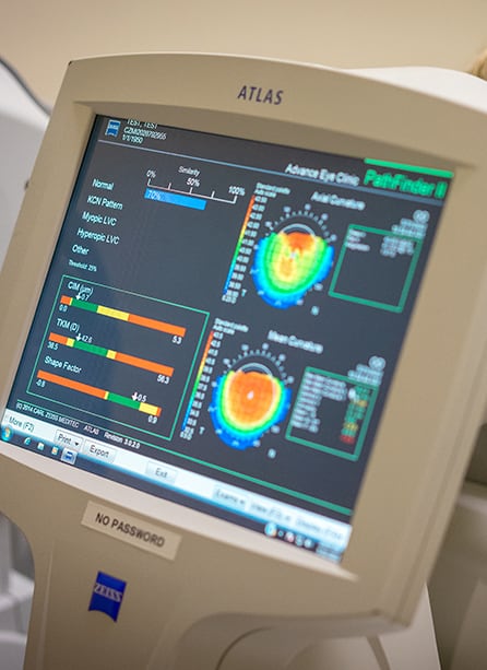
- Keratoconus (deterioration of the cornea, leading to change of shape and loss of vision)

Corneal Diseases
Cornea conditions can be caused by multiple factors, from injuries to lifestyle changes. Therefore, regular eye exams are essential for managing cornea health.
It’s also recommended to talk with your eye care provider when you experience eye symptoms, such as eye discomfort or vision changes. Read more about common cornea conditions and their symptoms to discover proper corneal treatment.
Scleritis
Scleritis, or inflammation of the sclera, can present as a painful red eye with or without vision loss. There are two type of scleritis:
- Anterior scleritis, the most common form, is characterized by severe pain and extreme scleral tenderness. The sclera is notably white, avascular and thin.
- Posterior scleritis is defined as involvement of the sclera posterior to the insertion of the rectus muscles. Posterior scleritis, although rare, can manifest as serious retinal detachment, choroidal folds, or both. There is often a loss of vision as well as pain upon eye movement.
Keratitis
Keratitis is a condition where the cornea—the clear, round dome covering the eye’s iris and pupil—becomes swollen or inflamed, making the eye red and painful and affecting vision. Keratitis is also known as a corneal ulcer.
Some forms of keratitis may involve infection, including bacterial keratitis, viral keratitis, fungal keratitis, and parasitic keratitis.
Contact lens wearers need to remember that infectious keratitis can result from not caring for your contact lenses properly.
A non-infectious form of keratitis may be caused by a simple fingernail scratch or from wearing your contact lenses too long. Whatever form of keratitis you may have, it is crucial that you see an ophthalmologist right away. Waiting to have your keratitis diagnosed and treated can lead to serious complications, including blindness.
Keratitis or corneal ulcer causes can include:
- Bacterial infection
- Viral infection
- Fungal infection (from plant material in the eye)
- Parasitic infection
- Improper cleaning and/or care for contact lenses
- Wearing contact lenses too long
- Injury (scratch)
- Vitamin A deficiency (rare)
Keratitis or corneal ulcer treatment depends on the type and severity of this corneal problem. Antibacterial or antifungal eye drops may be used to treat corneal infections. Sometimes steroid eye drops may be necessary to reduce the inflammation (swelling) of keratitis.
If the cornea is severely scarred or thinning has occurred, a corneal transplant may be needed to restore vision.
It is important to remember that keratitis must be treated early to reduce the risk of complications, and it is likely that frequent visits to an ophthalmologist may be needed to fully treat this corneal problem.
Corneal Ulcers
A corneal ulcer is an open sore in the surface layer of the eye, typically caused by a corneal infection or cornea swelling (keratitis). Keratitis can be infectious (bacterial, viral, or fungal) or non-infectious (eye injury). Chronic dry eye and unsterilized contact lenses are also risk factors.
In addition to symptoms caused by keratitis, corneal ulcers can also cause:
- Blurry vision
- Eye discharge or pus
- Eye inflammation
- Eyelid swelling
- Eye soreness
- Feeling something stuck in the eye
- Light sensitivity
- Watery eyes
- White spot on the cornea
A corneal ulcer can be treated with antibacterial, antiviral, or antifungal eye medication. By treating the infection, swelling may reduce, allowing the sore to heal. An eye doctor may also prescribe corticosteroid eye drops to help with inflammation.
In severe cases, a corneal transplant may be necessary to remove the damaged tissue.
Fuchs’ Dystrophy
Fuchs’ dystrophy is a disease of the cornea. It is when cells in the corneal layer called the endothelium die off. These cells normally pump fluid from the cornea to keep it clear. When they die, fluid builds up and the cornea gets swollen and puffy. Vision becomes cloudy or hazy.
Fuchs’ dystrophy has two stages. In the early stage (stage 1), you may notice few, if any, problems. In the later stage (stage 2), your blurry or hazy vision will not get better as the day goes on. Here are other symptoms:
- Sandy or gritty feeling in your eyes
- Eye pain from the tiny blisters in the cell layer of the cornea
- Being extra sensitive to bright light
- Eye problems get worse in humid areas
- Very blurry or hazy vision from scarring at the center of the cornea
To diagnose these conditions, your ophthalmologist will look closely at your cornea and measure its thickness. This is called pachymetry. They will also check for tiny blisters. Using a special photograph of your cornea, your ophthalmologist may count the cells in your endothelium.
There is no cure for Fuchs’ dystrophy. However, you can control vision problems from corneal swelling. Your treatment depends on how Fuchs’ dystrophy affects your eye’s cells, and may include:
- Eye Drop medicine or ointment to reduce swelling of the cornea’s cells.
- Endothelial keratoplasty (EK): Healthy endothelial cells are transplanted into your cornea.
- Full corneal transplant: The center of your cornea is replaced with a healthy donor cornea.
Your ophthalmologist will discuss what treatments are best for your condition.
Keratoconus
Keratoconus is a vision disorder resulting from a cornea that is too thin or irregularly shaped. The “conus” part of the name comes from the cone-like shape that can occur. The abnormal shape prevents the cornea from focusing light correctly.
In the early stages of keratoconus, symptoms may include slightly blurry vision and increased light sensitivity. As the disorder develops, the cornea bulges and symptoms can increase significantly. In some cases, the cornea can swell and crack. Although the crack can heal, it becomes scar tissue which can interfere with cornea function. Swelling can also continue without treatment.
When symptoms appear can depend on the cause and when the disorder began. It can commonly develop in the late teens or early 20s, typically progressing for 10–20 years before slowing. Keratoconus can be inherited, but frequent eye rubbing is also a risk factor.
Contact lenses and contact lenses can help correct mild astigmatism or myopia caused by keratoconus. In later stages of development, rigid gas permeable (RGP) contact lenses or surgery may be necessary to preserve vision.
Corneal cross-linking treatment can strengthen or reinforce the cornea. It can slow progression, but cannot reverse corneal bulging or thinning. Another option is a corneal transplant surgery, which replaces the cornea or a portion of the cornea.
Dry Eye
Dry eye is a common condition affecting tear production or quality. When tears work effectively, they protect, nourish, and lubricate the eye’s surface. When your eyes don’t produce enough tears or lack necessary components, it can result in:
- Blurry vision
- Eye redness
- Gritty or scratchy eyes
- Light sensitivity
- Stringy mucus
- Watery eyes
Untreated chronic dry eye can cause cornea inflammation and damage, leading to impaired vision. Dry eye therapy can help patients find relief and help protect the cornea.
Corneal Transplant Surgery
The cornea is the clear, front window of the eye. It helps focus light into the eye so that you can see. The cornea is made of layers of cells, and these layers work together to protect your eye and provide clear vision.
Your cornea must be clear, smooth and healthy for good vision. If it is scarred, swollen, or damaged, light is not focused properly into the eye. As a result, your vision is blurry or you see glare. If your cornea cannot be healed or repaired, your ophthalmologist may recommend a corneal transplant. This is when the diseased cornea is replaced with a clear, healthy cornea from a human donor.
There are different types of corneal transplants. In some cases, only the front and middle layers of the cornea are replaced. In others, only the inner layer is removed. Sometimes, the entire cornea needs to be replaced.
Full Thickness Cornea Transplant
Your entire cornea may need to be replaced if both the front and inner corneal layers are damaged. This is called penetrating keratoplasty (PK), or full thickness cornea transplant. Your diseased or damaged cornea is removed. Then the clear donor cornea is sewn into place.
PK has a longer recovery period than other types of cornea transplants. Getting complete vision back after PK may take up to 1 year or longer.
With a PK, there is a slightly higher risk than with other types of corneal transplants that the cornea will be rejected. This is when the body’s immune system attacks the new corneal tissue.
Partial Thickness Corneal Transplant
Sometimes the front and middle layers of the cornea are damaged. In this case, only those layers are removed. The endothelial layer, or the thin back layer, is kept in place. This transplant is called deep anterior lamellar keratoplasty (DALK) or partial thickness corneal transplant. DALK is commonly used to treat keratoconus or bulging of the cornea.
Healing time after DALK is shorter than after a full cornea transplant. There is also less risk of having the new cornea rejected.
Endothelial Keratoplasty
When the innermost layer of the cornea (the endothelium) is damaged, an endothelial keratoplasty removes the layer. Then, the layer is replaced with healthy donor tissue. Depending on the position and endothelium thickness, the surgeon may perform a DSEK (DSAEK) or DMEK procedure.
Full recovery after endothelial keratoplasty can take up to 6 months, but patients may resume most normal activities within weeks.
Corneal Cross-Linking
“Keratoconus” or “ectasia” is progressive thinning and weakening of the cornea, which is the outer-most layer of the eye. This progressive condition can lead to permanently
decreased vision, require a corneal transplant, or even perforation of the eye if it is not treated.
Corneal collagen cross-linking is the only FDA-approved, minimally invasive treatment for keratoconus and post-LASIK ectasia that combines riboflavin (vitamin B2) drops and UV light in a chemical reaction that strengthens corneal fibers and prevents worsening of ectasia. This procedure takes around 60 minutes to complete.
If cross-linking is performed early on in the disease process, the shape of the cornea can be stabilized, halting the progression of this disease and ultimately preventing the visual acuity from worsening. It is important to treat unstable keratoconus as early as possible to prevent complications or the need for corneal
transplantation in the future.
Locations
We have several convenient locations throughout South Carolina. Please view the nearest location to you or get directions below.
Our Services
News
How to Reduce the Risk of Eye Problems in the Workplace
Dry EyeReviewed By: Dr. Kimberly Carter, OD In the hustle of modern workplaces, our eyes work overtime—be it glued to screens, navigating bustling offices, or handling tools and chemicals. Yet, we often overlook the strain until it manifests as constant rubbing, headaches, or blurred vision. Eye problems at work aren’t just about accidents or dramatic injuries. […]
Read More… from How to Reduce the Risk of Eye Problems in the Workplace
New Laser Eye Treatment Available for Glaucoma Patients in the Lowcountry
Glaucoma, NewsCarolina Eyecare Physicians is proud to offer the recently released Voyager™ DSLT laser treatment for patients with glaucoma. The practice is among the first in the country to utilize this technology to perform Direct Selective Laser Trabeculoplasty (DSLT). Additionally, the procedure is the first of its kind to be offered in the state of South […]
Read More… from New Laser Eye Treatment Available for Glaucoma Patients in the Lowcountry
The Role of Eye Exams in Detecting Early Signs of Age-Related Macular Degeneration (AMD)
Age-Related Macular DegenerationReviewed By: Dr. Jonathan Brugger, MD Imagine waking up one day and realizing the center of your vision is blurred—faces look hazy, reading becomes a struggle, and everyday tasks feel harder. This is how macular degeneration can impact your life and often creeping in silently until it starts affects your life. But here’s the good […]
How to Reduce the Risk of Eye Problems in the Workplace

Reviewed By: Dr. Kimberly Carter, OD In the hustle of modern workplaces, our eyes work overtime—be it glued to screens, navigating bustling offices, or handling tools and chemicals. Yet, we often overlook the strain until it manifests as constant rubbing, headaches, or blurred vision. Eye problems at work aren’t just about accidents or dramatic injuries. […]
Read More… from How to Reduce the Risk of Eye Problems in the Workplace
New Laser Eye Treatment Available for Glaucoma Patients in the Lowcountry

Carolina Eyecare Physicians is proud to offer the recently released Voyager™ DSLT laser treatment for patients with glaucoma. The practice is among the first in the country to utilize this technology to perform Direct Selective Laser Trabeculoplasty (DSLT). Additionally, the procedure is the first of its kind to be offered in the state of South […]
Read More… from New Laser Eye Treatment Available for Glaucoma Patients in the Lowcountry
The Role of Eye Exams in Detecting Early Signs of Age-Related Macular Degeneration (AMD)

Reviewed By: Dr. Jonathan Brugger, MD Imagine waking up one day and realizing the center of your vision is blurred—faces look hazy, reading becomes a struggle, and everyday tasks feel harder. This is how macular degeneration can impact your life and often creeping in silently until it starts affects your life. But here’s the good […]


We are a proud partner of US Eye, a leading group of patient-centric, vertically integrated multi-specialty physician practices providing patients with care in ophthalmology, optometry, dermatology, audiology, and cosmetic facial surgery.


